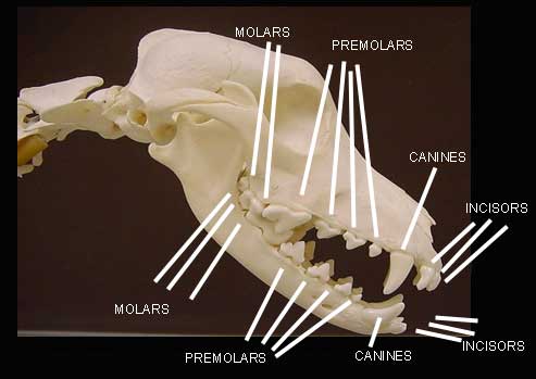
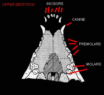
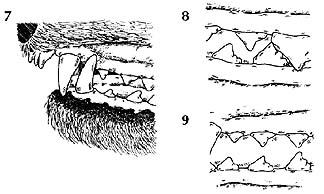
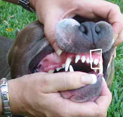
| The incisor teeth are the 6 small teeth located at the center of each dental arch. Next are the canine or "fang" teeth. All the teeth behind the canine teeth are referred to as cheek teeth. In the permanent dentition, there are 4 premolars on each side in each jaw. The upper fourth premolars are the largest cutting teeth of the upper jaw. They are referred to as carnasial teeth or "shearing" teeth. Finally, the molars are the large grinding teeth in the back of the mouth.
When examining or showing a dog's dentition, be sure to clear the lips and cheek tissue clearly from the mouth or the teeth may be miscounted or misidentified. Be sure the dog is not nervous and setting its teeth in an abnormal position due to tenseness. Try to "soften" the jaw by relaxing the dog's head and neck position. Tooth placement is affected by genetics, jaw structure, lip structure, the tongue, other teeth - deciduous ("baby teeth") and/or permanent, as well as the habits of the dog such as carrying a training dummy, chewing on kennel fencing or crate wire, or continual rock fetching.
When evaluating dog's teeth, we need to look at the relationship of all the teeth to each other and the jaw. Ethical breeders do care about dentition and require knowledge about the whole mouth - not just the incisors. It is very easy to just count the number of teeth or evaluate the "bite" of the incisors, but it is only when we look at the overall picture that we can see how genetics is affecting the dentition. The breeder needs to know the number of teeth; the type of bite or how the incisors meet; the relationship of the canine teeth, the premolars, the molars, and the jaw curvature. If there are any genetic inconsistencies, this should be taken into consideration in the breeding program.
Here is a step-by-step method of evaluating the teeth (FROM Holstrom, Frost, and Gammon1992 Veterinary Dental Techniques W.B. Saunders Company)
Step 1 Observe the symmetry of the head, face, and teeth.
Step 2 Count the teeth. The teeth are divided into 4 quadrants. Upper and lower left; Upper and lower right. There are 3 upper left incisors; 1 upper left canine; 4 upper left premolars; 2 upper left molars. It is the same for the upper right teeth. There are 3 lower left incisors; 1 lower left canine; 4 lower left premolars; 3 lower left molars . It is the same for the lower right teeth. Total teeth in an adult canine should be 42.
Step 3 Evaluate the incisors. The normal incisor occlusion has the large cusp ( a pointed or rounded protuberance making up a divisional part of the chewing surface of a tooth) of the lower incisors occluding near the cingulum (the lingual lobe of an anterior tooth; lingual refers to the surface of the tooth facing the tongue) on the lingual side of the upper incisors. The large cusps of the central incisors should be centered with each other. The second and third incisors lose their centered orientation and the large cusp of the third incisor should be in the interproximal space (space between adjoining teeth) between the second and third maxillary incisors (maxillary refers to the upper jaw). All the incisors should be in an evenly curved line with no rotation. Rotation would mean that the tooth is not seated properly on the jaw bone. The axis of the tooth should be parallel to the jaw.
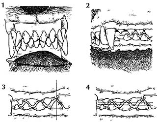
Step 4 Observe the relationship of the canine teeth. The mandibular canine or "fang" tooth should occlude buccal (toward the cheek tissue) to the gingiva of the maxilla and should divide the space between the maxillary canine tooth and the maxillary third incisor. This is the most reliable reference point in the mouth.
Step 5 Observe the relationship of the premolars. The large cusp on the lower fourth premolar should divide the space between the upper third and fourth premolars.
Step 6 Observe the occlusal plane of the upper and lower arches. The occlusal surface is the surface of the tooth that faces the opposite dental arches' chewing surface. The premolars should interdigitate from the second premolars back to the cusps of the upper fourth premolar with overlapping of the cusp tips. The molars should occlude to allow the cusps to function in crushing. The premolars and molars should be aligned mesial (toward the center line of the dental arch) to distal ( position on the dental arch farther from the median line of the jaw) in a slightly curved line with none of the teeth rotated.
After evaluating the teeth and their relationship to the jaw, the dentition can be categorized into the following types of occlusions. The normal occlusion is a "scissor bite." This is the pattern in which the lower incisors occlude next to the cingulum (lingual lobe of an anterior tooth) on the lingual surface of the upper incisors.
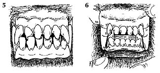
Dogs with a Class 1 occlusion have a normal occlusion with one or more teeth out of alignment or rotated. The following bites are Class 1: 1) the anterior crossbite where one or more of the lower incisors are anterior (situated in front of) to the upper incisors and the rest of the teeth occlude normally; 2) the level bite where the upper and lower incisors occlude cusp to cusp ("butt bite"); and 3) the base narrow bite where the tips of the mandibular canine teeth are displaced lingually (toward the tongue) and occlude on the hard palate.
Dogs with a Class 2 occlusion have the lower premolars and molars positioned behind the normal relationship. This occlusion may also be termed brachygnathism, overshot, "parrot mouth," retrusive mandible, or distal mandibular excursion.
Dogs with a Class 3 occlusion have the lower premolars and molars positioned ahead (anterior) of the normal relationship. This occlusion may also be termed prognathism; undershot; "Bulldog bite"; protrusive mandible; or mesial mandibular excursion.
An unclassified bite is the "wry bite." An abnormal occlusion caused by a difference in length of the two maxillae (upper jaw bones) and the mandibles (lower jaw bones). The abnormal occlusion results in a variety of different jaw relationships as one side of the jaw grows faster than the other and distorts or "twists" the mouth giving it a "wry" appearance. This condition is quite a handicap and leads to difficulty in grasping and chewing food as well as retrieving game.
It is only when we as dog handlers, breeders, and/or judges evaluate the dog's entire mouth that we can effectively understand how genetics and the environment are affecting the dog's dentition. Each dog should have a good oral exam to determine any health concerns or breeding considerations.
~Versatile Hunting Dog Magazine, December 2001
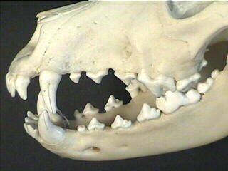
We can see from this image that these teeth have the proper occlusion. Imagine as the dog is closing its mouth the teeth will meet each other in a nice mesh!
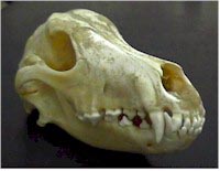
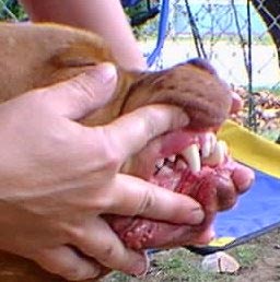
These teeth do not mesh quite right. The bite is a bit flush and the pre-molars P2 are almost on top of each other. The canines however are in the correct place. This is not the worse fault for the APBT. This dog could still function well at that task for which it was intended. This is ONLY a Fault not a serious fault.
|

